Quantcast
Channel: Cancer – www.thctotalhealthcare.com
Viewing all 779 articles
Browse latest View live

Management of chronic pain with Jalaprakshalana (water-wash) Shodhita (processed) Bhanga ( Cannabis sativa L.) in cancer patients with deprived quality of life: An open-label single arm clinical trial

$
0
0

“Introduction: Pain is a common and complex symptom of cancer having physical, social, spiritual and psychological aspects. Approximately 70%-80% of cancer patients experiences pain, as reported in India. Ayurveda recommends use of Shodhita (Processed) Bhanga (Cannabis) for the management of pain but no research yet carried out on its clinical effectiveness.

Objective: To assess the analgesic potential of Jala-Prakshalana (Water-wash) processed Cannabis sativa L. leaves powder in cancer patients with deprived quality of life (QOL) through openlabel single arm clinical trial.

Materials and methods: Waterwash processed Cannabis leaves powder filled in capsule, was administered in 24 cancer patients with deprived QOL presenting complaints of pain, anxiety or depression; for a period of 4 weeks; in a dose of 250 mg thrice a day; along with 50 ml of cow’s milk and 4 g of crystal sugar. Primary outcome i.e. pain was measured by Wong-Bakers FACES Pain Scale (FACES), Objective Pain Assessment (OPA) scale and Neuropathic Pain Scale (NPS). Secondary outcome namely anxiety was quantified by Hospital Anxiety and Depression Scale (HADS), QOL by FACT-G scale, performance score by Eastern Cooperative Oncology Group (ECOG) and Karnofsky score.

Results: Significant reduction in pain was found on FACES Pain Scale (P < 0.05), OPA (P < 0.05), NPS (P < 0.001), HADS (P < 0.001), FACT-G scale (P < 0.001), performance status score like ECOG (P < 0.05) and Karnofsky score (P < 0.01).

Conclusion: Jalaprakshalana Shodhita Bhanga powder in a dose of 250 mg thrice per day; relieves cancerinduced pain, anxiety and depression significantly and does not cause any major adverse effect and withdrawal symptoms during trial period.”

https://pubmed.ncbi.nlm.nih.gov/31831967/

“Administration of Jalaprakshalana Shodhita Bhanga (water-wash processed Cannabis) leaves powder in dose of 250 mg thrice a day with 50 ml of cow’s milk and 4 g sugar as an adjuvant, for a period of 1 month; significantly relieves pain, anxiety and depression of cancer patients without creating any major side effects, dependency and withdrawal symptoms. Processed Cannabis is significantly effective for improvement in QOL of a cancer patient.”

https://www.ayujournal.org/article.asp?issn=0974-8520;year=2019;volume=40;issue=1;spage=34;epage=43;aulast=Tavhare


Cannabidiol exerts anti-proliferative activity via a cannabinoid receptor 2-dependent mechanism in human colorectal cancer cells

$
0
0
International Immunopharmacology

“Colorectal cancer is the third leading cause of cancer incidence and mortality in the United States. Cannabidiol (CBD), the second most abundant phytocannabinoid in Cannabis sativa, has potential use in cancer treatment on the basis of many studies showing its anti-cancer activity in diverse types of cancer, including colon cancer. However, its mechanism of action is not yet fully understood.

In the current study, we observed CBD to repress viability of different human colorectal cancer cells in a dose-dependent manner. CBD treatment led to G1-phase cell cycle arrest and an increased sub-G1 population (apoptotic cells); it also downregulated protein expression of cyclin D1, cyclin D3, cyclin-dependent kinase 2 (CDK2), CDK4, and CDK6. CBD further increased caspase 3/7 activity and cleaved poly(ADP-ribose) polymerase, and elevated expression of endoplasmic reticulum (ER) stress proteins including binding immunoglobulin protein (BiP), inositol-requiring enzyme 1α (IRE1α), phosphorylated eukaryotic initiation factor 2α (eIF2α), activating transcription factor 3 (ATF3), and ATF4.

We found that CBD repressed cell viability and induced apoptotic cell death through a mechanism dependent on cannabinoid receptor type 2 (CB2), but not on CB1, transient receptor potential vanilloid, or peroxisome proliferator-activated receptor gamma. Anti-proliferative activity was also observed for other non-psychoactive cannabinoid derivatives including cannabidivarin (CBDV), cannabigerol (CBG), cannabicyclol (CBL), and cannabigerovarin (CBGV). Our data indicate that CBD and its derivatives could be promising agents for the prevention of human colorectal cancer.”

https://pubmed.ncbi.nlm.nih.gov/35598400/

“CBD represses viability of human colorectal cancer cells.•

CBD induces cell cycle arrest and increases apoptosis and ER stress in human colorectal cancer cells.•

CBD represses cell viability and induces apoptotic cell death via a CB2-dependent mechanism.”

https://www.sciencedirect.com/science/article/pii/S1567576922003496?via%3Dihub


The Effectiveness and Safety of Medical Cannabis for Treating Cancer Related Symptoms in Oncology Patients

$
0
0
Frontiers in Pain Research (@FrontPain) / Twitter

“The use of medical cannabis (MC) to treat cancer-related symptoms is rising. However, there is a lack of long-term trials to assess the benefits and safety of MC treatment in this population. In this work, we followed up prospectively and longitudinally on the effectiveness and safety of MC treatment.

Oncology patients reported on multiple symptoms before and after MC treatment initiation at one-, three-, and 6-month follow-ups. Oncologists reported on the patients’ disease characteristics. Intention-to-treat models were used to assess changes in outcomes from baseline. MC treatment was initiated by 324 patients and 212, 158 and 126 reported at follow-ups.

Most outcome measures improved significantly during MC treatment for most patients (p < 0.005). Specifically, at 6 months, total cancer symptoms burden declined from baseline by a median of 18%, from 122 (82–157) at baseline to 89 (45–138) at endpoint (−18.98; 95%CI= −26.95 to −11.00; p < 0.001). Reported adverse effects were common but mostly non-serious and remained stable during MC treatment.

The results of this study suggest that MC treatment is generally safe for oncology patients and can potentially reduce the burden of associated symptoms with no serious MC-related adverse effects.

The main finding of the current study is that most cancer comorbid symptoms improved significantly during 6 months of MC treatment.

Additionally, we found that MC treatment in cancer patients was well tolerated and safe.”

https://www.frontiersin.org/articles/10.3389/fpain.2022.861037/full?utm_source=fweb


Cannabinoid Receptor-1 suppresses M2 macrophage polarization in colorectal cancer by downregulating EGFR

$
0
0
Cell Death Discovery

“Cannabinoid receptors, CB1 and CB2, have been implicated as emerging targets for cancer therapy. Herein, we investigated the potential regulation mechanism of CB1 and its implications in colorectal cancer. CB1 and EGFR expression were examined in colorectal cancer cell lines. The effects of CB1 agonist ACEA and its antagonist AM251 on the proliferation, migration and invasion of colorectal cancer cells and the expression of M1 and M2 macrophage markers were examined. EGFR overexpression was performed with plasmids containing EGFR gene. Tumor xenografts were constructed to explore the effects of CB1 activation on tumorigenesis. We showed that CB1 was downregulated while EGFR was upregulated in colorectal cancer cells. The activation of CB1 suppressed the proliferation, migration and invasion of colorectal cancer cells and the differentiation of M2 macrophages, while CB1 inhibition had opposite effects. Moreover, the alterations in tumorigenesis and M2 macrophage activation induced by CB1 activation were counteracted by EGFR overexpression. Besides, CB1 silencing promoted tumor cell proliferation and M2 polarization which was counteracted by EGFR knockdown. In vivo, CB1 activation also repressed tumorigenesis and M2 macrophage activation. The present study demonstrated that CB1 activation suppressed M2 macrophage through EGFR downregulation in colorectal cancers. These findings first unveiled the potential avenue of CB1 as a targeted therapy for colorectal cancer.”

https://pubmed.ncbi.nlm.nih.gov/35641479/

“In conclusion, our study showed that CB1 activation suppressed tumor growth and M2 macrophage activation in colorectal cancer by downregulating EGFR. Our study provided the first evidence that CB1 activation was capable to suppress M2 macrophage activation. Since M2 macrophage are linked with immune evasion in various cancers, CB1 might be a promising target for cancer treatment.”

https://www.nature.com/articles/s41420-022-01064-8

“The use of THC, or selective targeting of the CB1 receptor, may represent a novel strategy for colorectal cancer therapy.”

https://pubmed.ncbi.nlm.nih.gov/17583570/

Biological Effects of Cannabidiol on Human Cancer Cells: Systematic Review of the Literature

$
0
0
Pharmacological Research


“This systematic review examine the biological effects of CBD, a major component of therapeutic Cannabis, on human pathological and cancer cell populations of integumentary, gastro-intestinal, genital and breast, respiratory, nervous, haematopoietic and skeletal districts in terms of cell viability, proliferation, migration, apoptosis, inflammation, metastasis, and CBD receptor expression. The included studies were in English, on human cell lines and primary culture from non-healthy donors with CBD exposure as variable and no CBD exposure as control. Quality assessment was based on ToxRtool with a reliability score ranging from 15 to 18. Following the PRISMA statement 4 independent reviewers performed an electronic search using MEDLINE via PubMed, Scopus and Web of Science. From 3974 articles, 83 studies have been selected. Data showed conflicting results due to different concentration exposure, administrations and time points. CBD inhibited cell viability and proliferation in most cellular districts except the integumentary apparatus. Also a significant inhibition of migration was observed in all cell types, while an increase in apoptosis at both high and low doses (greater and less than 10μM respectively). Considering inflammation, CBD caused an anti-inflammatory effect on nervous cells at low doses and on gastro-intestinal cells at high doses, while metastatic power was reduced even at low doses, but in a skeletal cell line there was an increased angiogenesis. CB1 receptor has been related to viability effects, CB2 to apoptosis and TRPV1 to inflammation and invasiveness. A detailed insight into these aspects would allow therapeutic use of this substance without possible side effects.”

https://pubmed.ncbi.nlm.nih.gov/35643249/

https://www.sciencedirect.com/science/article/abs/pii/S1043661822002122?via%3Dihub


Protective Effects of Cannabidiol on Chemotherapy-Induced Oral Mucositis via the Nrf2/Keap1/ARE Signaling Pathways

$
0
0
Archive of "Oxidative Medicine and Cellular Longevity". - PMC

“Oral mucositis (OM) is a common complication during chemotherapy characterized by ulceration, mucosa atrophy, and necrosis, which seriously interferes with nutritional intake and oncotherapy procedures among patients. However, the efficacy of current treatments for OM remains limited.

Cannabidiol (CBD) is a natural cannabinoid with multiple biological activities, including antioxidant and anti-inflammatory potential. In this study, we aimed to investigate the chemopreventive effects and mechanisms of CBD in protecting C57BL/6N mice and human oral keratinocytes (HOK) from 5-fluorouracil- (5-FU-) induced OM.

Here, we found that CBD alleviated the severity of 5-FU-induced OM in mice, including improved survival, decreased body weight loss, reduced ulcer sizes, and improved clinical scores. Histologically, CBD restored epithelial thickness and normal structure in tongue tissues. Meanwhile, CBD attenuated reactive oxygen species (ROS) overproduction and improved the antioxidant response, suppressed the inflammatory response, promoted the proliferation of epithelial cells, and inhibited 5-FU-induced apoptosis. In vitro, consistent outcomes showed that CBD suppressed cellular ROS levels, enhanced antioxidant ability, reduced inflammatory response, promoted proliferation, and inhibited apoptosis in 5-FU-treated HOK cells. In particular, CBD upregulated the expression levels of antioxidant enzymes, heme oxygenase-1 (HO-1) and NAD(P)H quinine oxidoreductase 1 (NQO1), by increasing the expression and nuclear translocation of nuclear factor erythroid 2-related factor 2 (Nrf2) and decreasing Kelch-like ECH-associated protein 1 (Keap1). Notably, the Nrf2 inhibitor ML385 reversed the protective effect of CBD. Nrf2-siRNA transfection also significantly blunted the antioxidant effect of CBD in in vitro OM model.

Collectively, our findings suggested that CBD protected against 5-FU-induced OM injury at least partially via the Nrf2/Keap1/ARE signaling pathways, highlighting the therapeutic prospects of CBD as a novel strategy for chemotherapy-induced OM.”

https://pubmed.ncbi.nlm.nih.gov/35669853/

“CBD alleviates chemotherapy-induced OM and protects against the toxicity of 5-FU by improving oxidative stress defense, downregulating mucosal inflammation, promoting cell proliferation, and inhibiting 5-FU-induced apoptosis both in mice and in HOK. Moreover, CBD-activated Nrf2/Keap1/ARE signaling pathways might be the underlying mechanism for OM recovery.”

https://www.hindawi.com/journals/omcl/2022/4619760/


Extracellular vesicles of cannabis with high CBD content induce anticancer signaling in human hepatocellular carcinoma

$
0
0
Biomedicine & Pharmacotherapy


“Plant-derived extracellular vesicles (EVs) have been the topic of interest in recent years due to their proven therapeutic properties. Intact or manipulated plant EVs have shown antioxidant, anti-inflammatory, and anti-cancerous activities as a result of containing bioactive metabolites and other endogenous molecules. Less is known about the EV efficacy with high levels of bioactive secondary metabolites derived from medicinal or non-edible plants.

Numerous data suggest the functionality of Cannabis sativa extract and its phytocannabinoids in cancer treatment. Here, two chemotypes of cannabis with different levels of D-9-tetrahydrocannabinol (THC) and cannabidiol (CBD) were selected. EVs were isolated from each chemotype via differential ultracentrifugation. HPLC analysis was illustrative of the absence of THC in EVs derived from both plants. Therefore, two types of EVs were classified according to their CBD content into high- (H.C-EVs) and low-CBD EVs (L.C-EVs). Electron microscopy and DLS showed both cannabis-derived EVs (CDEVs) can be considered as exosome-like nanovesicles. Cytotoxicity assay showed that H.C-EVs strongly decreased the viability of two hepatocellular carcinoma (HCC) cell lines, HepG2 and Huh-7, in a dose and time-dependent manner compared with L.C-EVs. H.C-EVs had no significant effect on HUVECs normal cell growth. The finding showed that the H.C-EVs arrested the G0/G1 phase in the cell cycle and significantly induced cell death by activating mitochondrial-dependent apoptosis signaling pathways in both HCC cell lines.

Altogether, the current study highlights that CDEVs can be an ideal natural vehicle for bioactive phytocannabinoids and a promising strategy in cancer management.”

https://pubmed.ncbi.nlm.nih.gov/35667235/

“Altogether, our findings suggest that the EVs derived from cannabis can act as natural nano-carriers containing bioactive phytochemicals and be used in cancer research. The possible use of these biomaterials in combination with chemotherapy drugs can open a new gateway for cancer treatment.”

https://www.sciencedirect.com/science/article/pii/S0753332222005984?via%3Dihub

Fig. 1

Cannabidiol and Cannabigerol Inhibit Cholangiocarcinoma Growth In Vitro via Divergent Cell Death Pathways

$
0
0
biomolecules-logo

“Cholangiocarcinoma (CCA) is a rare and highly lethal disease with few effective treatment options.

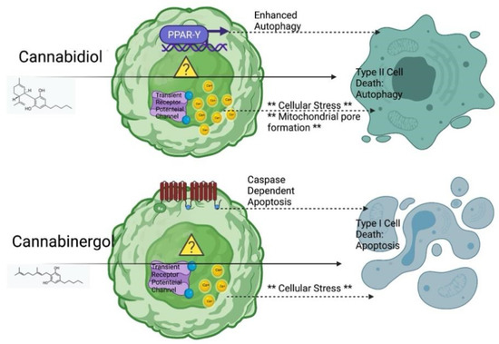
Cannabinoids, cannabidiol (CBD) and cannabigerol (CBG) are non-psychedelic components extracted from cannabis. These non-psychoactive compounds have shown anti-proliferative potential in other tumor models; however, the efficacy of CBD and CBG in CCA is unknown. Furthermore, two cell death pathways are implicated with CBD resulting in autophagic degeneration and CBG in apoptosis. HuCC-T1 cells, Mz-ChA-1 cells (CCA cell lines) and H69 cells (immortalized cholangiocytes), were treated with CBD and CBG for 24 to 48 h.

The influence of these cannabinoids on proliferation was assessed via MTT assay. Apoptosis and cell cycle were evaluated via Annexin-V apoptosis assay and propidium iodide, respectively. The expression of proliferation biomarker Ki-67, apoptosis biomarker BAX, and autophagic flux biomarkers LC3b and LAMP1 were evaluated via immunofluorescence. Cell migration and invasion were evaluated via wound healing assay and trans-well migration invasion assays, respectively. The colony formation was evaluated via colony formation assay. In addition, the expression of autophagy gene LC3b and apoptosis genes BAX, Bcl-2, and cleaved caspase-3 were evaluated via Western blot.

CBD and CBG are non-selective anti-proliferative agents yielding similar growth curves in CCA; both cannabinoids are effective, yet CBG is more active at lower doses. Low doses of CBD and CBG enhanced immortalized cholangiocyte activity. The reduction in proliferation begins immediately and occurs maximally within 24 h of treatment. Moreover, a significant increase in the late-stage apoptosis and a reduction in the number of cells in S stage of the cell cycle indicates both CBD and CBG treatment could promote apoptosis and inhibit mitosis in CCA cells. The fluorescent expression of BAX and LC3b was significantly enhanced with CBD treatment when compared to control. LAMP1 and LC3b colocalization could also be observed with CBD and CBG treatment indicating changes in autophagic flux.

A significant inhibition of migration, invasion and colony formation ability was shown in both CBD and CBG treatment in CCA. Western blot showed an overall decrease in the ratio of anti-apoptotic protein Bcl-2 with respect to pro-apoptotic protein BAX with CBG treatment. Furthermore, CBD treatment enhanced the expression of Type II cell death (autophagic degeneration) protein LC3b, which was reduced in CBG-treated CCA cells. Meanwhile, CBG treatment upregulated Type I cell death (programmed apoptosis) protein cleaved caspase-3.

CBD and CBG are effective anti-cancer agents against CCA, capable of inhibiting the classic hallmarks of cancer, with a divergent mechanism of action (Type II or Type I respectively) in inducing these effects.”

https://pubmed.ncbi.nlm.nih.gov/35740979/

https://www.mdpi.com/2218-273X/12/6/854


Mechanisms of Cannabidiol (CBD) in Cancer Treatment: A Review

$
0
0
biology-logo


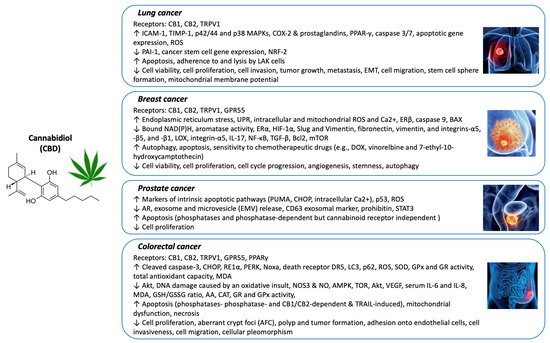
“Cannabis sativa L. (Cannabis) and its bioactive compounds, including cannabinoids and non-cannabinoids, have been extensively studied for their biological effects in recent decades. Cannabidiol (CBD), a major non-intoxicating cannabinoid in Cannabis, has emerged as a promising intervention for cancer research.

The purpose of this review is to provide insights into the relationship between CBD and cancer based on recent research findings.

The anticancer effects of CBD are mainly mediated via its interaction with the endocannabinoid system, resulting in the alleviation of pain and the promotion of immune regulation. Published reviews have focused on the applications of CBD in cancer pain management and the possible toxicological effects of its excessive consumption.

In this review, we aim to summarize the mechanisms of action underlying the anticancer activities of CBD against several common cancers. Studies on the efficacy and mechanisms of CBD on cancer prevention and intervention in experimental models (i.e., cell culture- and animal-based assays) and human clinical studies are included in this review.”

https://pubmed.ncbi.nlm.nih.gov/35741337/

“Emerging evidence suggests positive outcomes from the use of CBD as a cancer treatment. CBD can relieve cancer pain and ease the side effects of chemotherapy; however, there is less research about the mechanism of CBD’s anticancer effects. In this article, recent studies on the efficacy and mechanisms of CBD’s anticancer effects in cell- and animal-based models and human clinical studies are reviewed.”

https://www.mdpi.com/2079-7737/11/6/817


Cannabidiol Interacts Antagonistically with Cisplatin and Additively with Mitoxantrone in Various Melanoma Cell Lines-An Isobolographic Analysis

$
0
0
ijms-logo

“The medical application of cannabidiol (CBD) has been gathering increasing attention in recent years. This non-psychotropic cannabis-derived compound possesses antiepileptic, antipsychotic, anti-inflammatory and anxiolytic properties. Recent studies report that it also exerts antineoplastic effects in multiple types of cancers, including melanoma.

In this in vitro study we tried to reveal the anticancer properties of CBD in malignant melanoma cell lines (SK-MEL 28, A375, FM55P and FM55M2) administered alone, as well as in combination with mitoxantrone (MTX) or cisplatin (CDDP).

The effects of CBD on the viability of melanoma cells were measured by the MTT assay; cytotoxicity was determined in the LDH test and proliferation in the BrdU test. Moreover, the safety of CBD was tested in human keratinocytes (HaCaT) in LDH and MTT tests.

Results indicate that CBD reduces the viability and proliferation of melanoma-malignant cells and exerts additive interactions with MTX. Unfortunately, CBD produced antagonistic interaction when combined with CDDP. CBD does not cause significant cytotoxicity in HaCaT cell line.

In conclusion, CBD may be considered as a part of melanoma multi-drug therapy when combined with MTX. A special attention should be paid to the combination of CBD with CDDP due to the antagonistic interaction observed in the studied malignant melanoma cell lines.”

https://pubmed.ncbi.nlm.nih.gov/35743195/

https://www.mdpi.com/1422-0067/23/12/6752

In Vitro Effect of Δ9-Tetrahydrocannabinol and Cannabidiol on Cancer-Associated Fibroblasts Isolated from Lung Cancer

$
0
0
ijms-logo

“There is evidence that demonstrates the effect of cannabinoid agonists inhibiting relevant aspects in lung cancer, such as proliferation or epithelial-to-mesenchymal transition (EMT).

Most of these studies are based on evidence observed in in vitro models developed on cancer cell lines. These studies do not consider the complexity of the tumor microenvironment (TME). One of the main components of the TME is cancer-associated fibroblasts (CAFs), cells that are relevant in the control of proliferation and metastasis in lung cancer.

In this work, we evaluated the direct effects of two cannabinoid agonists, tetrahydrocannabinol (THC) and cannabidiol (CBD), used alone or in combination, on CAFs and non-tumor normal fibroblasts (NFs) isolated from adenocarcinoma or from healthy lung tissue from the same patients.

We observed that these compounds decrease cell density in vitro and inhibit the increase in the relative expression of type 1 collagen (COL1A1) and fibroblast-specific protein 1 (FSP1) induced by transforming growth factor beta (TGFβ). On the other hand, we studied whether THC and CBD could modulate the interactions between CAFs or NFs and cancer cells. We conditioned the culture medium with stromal cells treated or not with THC and/or CBD and cultured A549 cells with them.

We found that culture media conditioned with CAFs or NFs increased cell density, induced morphological changes consistent with EMT, inhibited cadherin-1 (CDH1) gene expression, and induced an increase in the relative expression of cadherin-2 (CDH2) and vimentin (VIM) genes in A549 cells. These changes were inhibited or decreased by THC and CBD administered alone or in combination. In another series of experiments, we conditioned culture media with A549 cells treated or not with THC and/or CBD, in the presence or absence of TGFβ. We observed that culture media conditioned with A549 in the presence of TGFβ induced an increase in the expression of COL1A1 and VIM, both in CAFs and in non-tumor NFs. Both THC and CBD ameliorated these effects.

In summary, the results presented here reinforce the usefulness of cannabinoid agonists for the treatment of some relevant aspects of lung cancer pathology, and demonstrate in a novel way their possible effects on CAFs as a result of their relationship with cancer cells. Likewise, the results reinforce the usefulness of the combined use of THC and CBD, which has important advantages in relation to the possibility of using lower doses, thus minimizing the psychoactive effects of THC.”

https://pubmed.ncbi.nlm.nih.gov/35743206/

https://www.mdpi.com/1422-0067/23/12/6766


Receptor-targeted nanoparticles modulate cannabinoid anticancer activity through delayed cell internalization

$
0
0
Scientific Reports

“Δ9-tetrahydrocannabinol (Δ9-THC) is known for its antitumor activity and palliative effects.

However, its unfavorable physicochemical and biopharmaceutical properties, including low bioavailability, psychotropic side effects and resistance mechanisms associated to dosing make mandatory the development of successful drug delivery systems.

In this work, transferring (Tf) surface-modified Δ9-THC-loaded poly(lactide-co-glycolic) nanoparticles (Tf-THC-PLGA NPs) were proposed and evaluated as novel THC-based anticancer therapy. Furthermore, in order to assess the interaction of both the nanocarrier and the loaded drug with cancer cells, a double-fluorescent strategy was applied, including the chemical conjugation of a dye to the nanoparticle polymer along with the encapsulation of either a lipophilic or a hydrophilic dye.

Tf-THC PLGA NPs exerted a cell viability decreased down to 17% vs. 88% of plain nanoparticles, while their internalization was significantly slower than plain nanoparticles. Uptake studies in the presence of inhibitors indicated that the nanoparticles were internalized through cholesterol-associated and clathrin-mediated mechanisms.

Overall, Tf-modification of PLGA NPs showed to be a highly promising approach for Δ9-THC-based antitumor therapies, potentially maximizing the amount of drug released in a sustained manner at the surface of cells bearing cannabinoid receptors.”

https://pubmed.ncbi.nlm.nih.gov/35079042/

“The potential therapeutic applications of marijuana, firstly reported in 1997 by the National Institutes of Health (NIH, USA), are attributed to a great extent to its main component, Δ9-tetrahydrocannabinol (Δ9-THC)1. This cannabinoid continues to attract special attention in oncology due to its palliative effects and antitumor activity; Δ9-THC has been reported to inhibit tumor angiogenesis and cell growth in malignant tissues, leading to cell death.”

“Δ9-THC has been reported to inhibit tumor angiogenesis and cell growth in malignant tissues.”

“Overall, Tf-modification of PLGA NPs seemed a highly promising approach for Δ9-THC-based antitumor therapies, aiming at a prolonged action of the carrier at the target cell surface. Moreover, the translation of this strategy to the delivery of alternative active pharmaceutical ingredients with pharmacological targets on the surface of cells could lead to advances in related therapies.”

https://www.nature.com/articles/s41598-022-05301-z

Cannabis: Chemistry, extraction and therapeutic applications

$
0
0
Chemosphere

“Cannabis, a genus of perennial indigenous plants is well known for its recreational and medicinal activities. Cannabis and its derivatives have potential therapeutic activities to treat epilepsy, anxiety, depression, tumors, cancer, Alzheimer’s disease, Parkinson’s disease, to name a few.

This article reviews some recent literature on the bioactive constituents of Cannabis, commonly known as phytocannabinoids, their interactions with the different cannabinoids and non-cannabinoid receptors as well as the significances of these interactions in treating various diseases and syndromes.

The biochemistry of some notable cannabinoids such as tetrahydrocannabinol, cannabidiol, cannabinol, cannabigerol, cannabichromene and their carboxylic acid derivatives is explained in the context of therapeutic activities.

The medicinal features of Cannabis-derived terpenes are elucidated for treating several neuro and non-neuro disorders. Different extraction techniques to recover cannabinoids are systematically discussed. Besides the medicinal activities, the traditional and recreational utilities of Cannabis and its derivatives are presented. A brief note on the legalization of Cannabis-derived products is provided.

This review provides comprehensive knowledge about the medicinal properties, recreational usage, extraction techniques, legalization and some prospects of cannabinoids and terpenes extracted from Cannabis.”

https://pubmed.ncbi.nlm.nih.gov/34838836/

“Cannabinoids have therapeutic effects against various health disorders.•

Medicinal effects are due to the interactions of cannabinoids with bio-receptors.•

Cannabinoids can be extracted from Cannabis plant products by eco-friendly extraction methods.”

https://www.sciencedirect.com/science/article/abs/pii/S0045653521034846?via%3Dihub

Image 1


Therapeutic Potential of Cannabinoids on Tumor Microenvironment: A Molecular Switch in Neoplasia Transformation

$
0
0

“The efficacy of chemotherapy depends on the tumor microenvironment. This microenvironment consists of a complex cellular network that can exert both stimulatory and inhibitory effects on tumor genesis.

Given the increasing interest in the effectiveness of cannabis, cannabinoids have gained much attention as a potential chemotherapy drug. Cannabinoids are a group of marker compounds found in Cannabis sativa L., more commonly known as marijuana, a psychoactive drug used since ancient times for pain management.

Although the anticancer potential of C. sativa, has been recognized previously, increased attention was generated after discovering the endocannabinoid system and the successful production of cannabinoid receptors.

In vitro and in vivo studies on various tumor models have shown therapeutic efficiency by modifying the tumor microenvironment.

This review summarizes the key literature surrounding the role of cannabinoids in the tumor microenvironment and their future promise in cancer treatment.”

https://pubmed.ncbi.nlm.nih.gov/35796303/

“Cannabis sativa L. is a natural source of valuable compounds that comprise cannabinoid agonists and antagonists, which have recently been scanned for future applications as anti-tumor drugs. Cannabinoids have mostly been used as a part of palliative care to alleviate pain, relieve nausea, and stimulate appetite in cancer patients. Although not yet approved for treating tumor progression, cannabinoid agonist/antagonists on the tumor microenvironment have been studied for the last 43 years. Research on cannabinoids and their potential therapeutic function has been ongoing since 1971. Numerous in vitro and in vivo studies have demonstrated the anti-cancer effects of cannabinoids in various cancer types.”

https://journals.sagepub.com/doi/10.1177/15347354221096766


The Combination of Δ 9-Tetrahydrocannabinol and Cannabidiol Suppresses Mitochondrial Respiration of Human Glioblastoma Cells via Downregulation of Specific Respiratory Chain Proteins

$
0
0
cancers-logo

“Phytocannabinoids represent a promising approach in glioblastoma therapy.

Previous work has shown that a combined treatment of glioblastoma cells with submaximal effective concentrations of psychoactive Δ9-tetrahydrocannabinol (THC) and non-psychoactive cannabidiol (CBD) greatly increases cell death.

In the present work, the glioblastoma cell lines U251MG and U138MG were used to investigate whether the combination of THC and CBD in a 1:1 ratio is associated with a disruption of cellular energy metabolism, and whether this is caused by affecting mitochondrial respiration.

Here, the combined administration of THC and CBD (2.5 µM each) led to an inhibition of oxygen consumption rate and energy metabolism. These effects were accompanied by morphological changes to the mitochondria, a release of mitochondrial cytochrome c into the cytosol and a marked reduction in subunits of electron transport chain complexes I (NDUFA9, NDUFB8) and IV (COX2, COX4). Experiments with receptor antagonists and inhibitors showed that the degradation of NDUFA9 occurred independently of the activation of the cannabinoid receptors CB1, CB2 and TRPV1 and of usual degradation processes mediated via autophagy or the proteasomal system.

In summary, the results describe a previously unknown mitochondria-targeting mechanism behind the toxic effect of THC and CBD on glioblastoma cells that should be considered in future cancer therapy, especially in combination strategies with other chemotherapeutics.”

https://pubmed.ncbi.nlm.nih.gov/35804909/

“Cannabidiol (CBD) is a phytocannabinoid from Cannabis sativa L. that exhibits no psychoactivity and, like the psychoactive cannabinoid Δ9-tetrahydrocannabinol (THC), shows anticancer effects in preclinical cell and animal models. Previous studies have indicated a stronger cancer-targeting effect when THC and CBD are combined. Here, we investigated how the combination of THC and CBD in a 1:1 ratio affects glioblastoma cell survival. The compounds were found to synergistically enhance cell death, which was attributed to mitochondrial damage and disruption of energy metabolism. A detailed look at the mitochondrial electron transfer chain showed that THC/CBD selectively decreased certain subunits of complexes I and IV. These data highlight the fundamental changes in cellular energy metabolism when cancer cells are exposed to a mixture of cannabinoids and underscore the potential of combining cannabinoids in cancer treatment.”

https://www.mdpi.com/2072-6694/14/13/3129


Investigating the Effects of a Synthetic Cannabinoid on the Pathogenesis of Leukemia and Leukemic Stem Cells: A New Therapeutic Approach

$
0
0
View details for Cannabis and Cannabinoid Research cover image

“The popularity and usage of synthetic cannabinoids (SCs) are increasing due to their easy accessibility and psychoactive effects worldwide. Studies on cannabinoids on leukemic stem cells (LSC) and hematopoietic stem cells (HSCs), which are the precursors of leukemia cells, generally depend on the natural cannabinoid delta-9-THC. As there is only a limited number of studies focusing on the results of SC applications, the reflections upon LSCs have to be clarified.

In this study, biological responses and antileukemic effects of JWH-018-one of the first produced and widely used SCs-were evaluated upon leukemia cells. Whether JWH-018 exhibited a preventive effect on both leukemic and HSCs was evaluated by presenting a therapeutic approach for the first time in the literature. Cells were analyzed in case of cell proliferation, apoptosis, and transcriptional expression profiling of some significant JAK/STAT and AKT/mTOR pathways, apoptotic, cell cycle regulation, and epigenetic chromatin remodeling-related genes following JWH-018 treatment.

In conclusion, however, further studies are still needed upon both HSCs and LSCs to illuminate the effects of SCs on leukemogenesis on chronic myeloid leukemia (CML) more clearly; we consider that the JWH-018 can provide a therapeutic effect on the pathogenesis of leukemia and particularly upon LSCs and SCs might have therapeutic potential in addition to current therapy.”

https://pubmed.ncbi.nlm.nih.gov/35834597/

https://www.liebertpub.com/doi/10.1089/can.2021.0180

“Dronabinol has preferential antileukemic activity in acute lymphoblastic and myeloid leukemia with lymphoid differentiation patterns. Our study provides rigorous data to support clinical evaluation of THC as a low-toxic therapy option in a well defined subset of acute leukemia patients.”

https://pubmed.ncbi.nlm.nih.gov/26775260/

“Cannabinoid CP55940 selectively induces apoptosis in Jurkat cells and in ex vivo T-cell acute lymphoblastic leukemia through H 2 O 2 signaling mechanism. Our findings support the use of cannabinoids as a potential treatment for T-ALL cells.”

https://pubmed.ncbi.nlm.nih.gov/32540572/

“CP 55,940 is a synthetic cannabinoid which mimics the effects of naturally occurring THC (one of the psychoactive compounds found in cannabis)”  https://en.wikipedia.org/wiki/CP_55,940

“Delta9-tetrahydrocannabinol-induced apoptosis in Jurkat leukemia T cells is regulated by translocation of Bad to mitochondria. Plant-derived cannabinoids, including Delta9-tetrahydrocannabinol (THC), induce apoptosis in leukemic cells”

https://pubmed.ncbi.nlm.nih.gov/16908594/


Protective Effects of Cannabis sativa on chemotherapy-induced nausea in a rat: Involvement of CB1 receptors

$
0
0

“Cyclophosphamide is an anticancer and immunosuppressive agent used in the treatment of various malignancies but causing gastrointestinal distress.

Cannabis sativa (C. sativa) and its derivatives have been used for the treatment of human gastrointestinal disorders. A purpose of this study was to investigate the effect of C. sativa on nausea induced by cyclophosphamide in rats.

Results showed that C. sativa ameliorates cyclophosphamide-induced emesis by increasing in body weight and normal diet intake with a decrease in kaolin diet intake after 7 days. Moreover, C. sativa significantly decreases (serotonin) 5HT, dopamine and noradrenaline, as well as, decreasing oxidative stress and inflammation. Administration of C. sativa significantly increased the expression of CB1R in intestinal homogenate. Treatment with C. sativa, also, improved the histological feature of an intestinal tissue.

These results suggested that C. sativa possess antiemetic, antioxidant and anti-inflammatory effects in chemotherapy-induced nausea in rats by activating CB1R.”

https://pubmed.ncbi.nlm.nih.gov/35861135/

https://onlinelibrary.wiley.com/doi/10.1111/fcp.12821

Anti-cancer properties of cannflavin A and potential synergistic effects with gemcitabine, cisplatin, and cannabinoids in bladder cancer

$
0
0

“Introduction: Several studies have shown anti-tumor effects of components present in cannabis in different models. Unfortunately, little is known about the potential anti-tumoral effects of most compounds present in cannabis in bladder cancer and how these compounds could potentially positively or negatively impact the actions of chemotherapeutic agents. Our study aims to evaluate the effects of a compound found in Cannabis sativa that has not been extensively studied to date, cannflavin A, in bladder cancer cell lines. We aimed to identify whether cannflavin A co-treatment with agents commonly used to treat bladder cancer, such as gemcitabine and cisplatin, is able to produce synergistic effects. We also evaluated whether co-treatment of cannflavin A with various cannabinoids could produce synergistic effects.

Results: Cell viability of bladder cancer cell lines was affected in a concentration-dependent fashion in response to cannflavin A, and its combination with gemcitabine or cisplatin induced differential responses-from antagonistic to additive-and synergism was also observed in some instances, depending on the concentrations and drugs used. Cannflavin A also activated apoptosis via caspase 3 cleavage and was able to reduce invasion by 50%. Interestingly, cannflavin A displayed synergistic properties with other cannabinoids like Δ9-tetrahydrocannabinol, cannabidiol, cannabichromene, and cannabivarin in the bladder cancer cell lines.

Discussion: Our results indicate that compounds from Cannabis sativa other than cannabinoids, like the flavonoid cannflavin A, can be cytotoxic to human bladder transitional carcinoma cells and that this compound can exert synergistic effects when combined with other agents. In vivo studies will be needed to confirm the activity of cannflavin A as a potential agent for bladder cancer treatment.”

https://pubmed.ncbi.nlm.nih.gov/35869542/

“A study recently demonstrated that the combination of Δ9-tetrahydrocannabinol and cannabichromene produced synergistic effects in a bladder cancer model, while another focused on the effects of cannabidiol and their potential formulation within nanoparticles to treat bladder cancer. Here, we show that other compounds from cannabis, like cannflavin A, may also induce beneficial cytotoxic and synergistic effects on bladder cancer cells. Our results also showed the ability of cannabinoids, other than Δ9-tetrahydrocannabinol, to produce synergistic effects when combined with the flavonoid cannflavin A.”

https://jcannabisresearch.biomedcentral.com/articles/10.1186/s42238-022-00151-y


Cannabidiol Regulates PPARγ-Dependent Vesicle Formation as well as Cell Death in A549 Human Lung Cancer Cells

$
0
0
pharmaceuticals-logo

“Extracts of phytocannabinoids from Cannabis sativa have been studied for therapeutic purposes. Although nonpsychoactive CBD has been studied as a promising anticancer drug because it induces apoptosis in many cancer cells, it is also known to induce several physiological changes.

In this study, we clarify the functional role it plays in the morphological characteristics of intracellular vesicle formation as well as apoptosis in A549 human lung cancer cells.

CBD treatment shows growth inhibition at concentrations above 20 μM, but FACS analysis shows low efficacy in terms of cell death. Microscopic observations suggest that multiple vesicles were detected in the cytoplasmic region of CBD-treated A549 cells.

CBD treatment upregulates apoptosis-related proteins, such as p53, PARP, RIP1, RIP3, Atg12, and Beclin, indicating that CBD regulates several types of cell death. CBD treatment also induced E-cadherin, PPARγ, clathrin, β-adaptin, and Tsg101, also known to be cellular-differentiation inducers or vesicle-formation components.

Treatment combining CBD with GW9662, a PPARγ inhibitor, reduced CBD-induced cytoplasmic vesicle formation. This indicates that PPARγ regulates the vesicle-formation mechanism. However, CBD-treated E-cad KO clones did not show this regulatory mechanism.

These results elucidate the pharmacological and molecular networks associated with CBD in PPARγ-dependent vesicle formation and the induction of apoptosis.”

https://pubmed.ncbi.nlm.nih.gov/35890134/

“Recently, the pharmacological efficacy of CBD has been focused on in many types of disease models. In this study, we have shown that CBD treatment upregulated cell death proteins, such as p53, PARP, RIP1, RIP3, Atg12, and Beclin. In addition, CBD treatment also induced E-cadherin, PPARγ, clathrin, β-adaptin, and Tsg101, also known to be cellular-differentiation inducers or vesicle-formation components.

We have also shown that PPARγ regulates the vesicle-formation mechanism. Therefore, not only have we confirmed cell death induction by CBD, we have also confirmed the regulation of vesicle formation by PPARγ in various cancer models.

Detailed molecuclar insights would enhance the therapeutic utilization of CBD without side effects. Cellular proliferation and death could be determined by a variety of molecular regulations in cellular networks composed of a variety of proteins. Moreover, the physiological changes in cells will be closely related to functional changes in intracellular organelles.

We suggest that an understanding of the complex intracellular network system is necessary to optimize the pharmacological efficacy of CBD. These steady research efforts will make significant progress in increasing the utilization of CBD, which has been restricted by legal regulations in some countries.”

https://www.mdpi.com/1424-8247/15/7/836/htm


Improved Therapeutic Efficacy of CBD with Good Tolerance in the Treatment of Breast Cancer through Nanoencapsulation and in Combination with 20(S)-Protopanaxadiol (PPD)

$
0
0
pharmaceutics-logo

“Cannabidiol (CBD), a nonpsychoactive major component derived from Cannabis sativa, widely used in neurodegenerative diseases, has now been proven to have growth inhibitory effects on many tumor cell lines, including breast tumors. Meanwhile CBD can effectively alleviate cancer-associated pain, anxiety, and depression, especially tumor cachexia, thus it is very promising as an anti-tumor drug with unique advantages.

20(S)-Protopanaxadiol (PPD) derived from the best-known tonic Chinese herbal medicine Ginseng was designed to be co-loaded with CBD into liposomes to examine their synergistic tumor-inhibitory effect. The CBD-PPD co-loading liposomes (CP-liposomes) presented a mean particle size of 138.8 nm. Further glycosyl-modified CP-liposomes (GMCP-liposomes) were prepared by the incorporation of n-Dodecyl β-D-maltoside (Mal) into the liposomal bilayer with glucose residue anchored on the surface to act as a ligand targeting the GLUT1 receptor highly expressed on tumor cells.

In vivo studies on murine breast tumor (4T1 cells)-bearing BALB/c mice demonstrated good dose dependent anti-tumor efficacy of CP-liposomes. A high tumor inhibition rate (TIR) of 82.2% was achieved with good tolerance. However, glycosylation modification failed to significantly enhance TIR of CP-liposomes.

In summary, combined therapy with PPD proved to be a promising strategy for CBD to be developed into a novel antitumor drug, with characteristics of effectiveness, good tolerance, and the potential to overcome tumor cachexia.”

https://pubmed.ncbi.nlm.nih.gov/35893789/

https://www.mdpi.com/1999-4923/14/8/1533/htm

Viewing all 779 articles
Browse latest View live